Foot Muscles Mri / Anatomy Of The Foot And Ankle Mri / The extrinsic muscles are located in the anterior and lateral compartments of the leg.
Get link
Facebook
X
Pinterest
Email
Other Apps
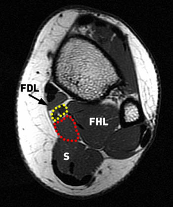
Foot Muscles Mri / Anatomy Of The Foot And Ankle Mri / The extrinsic muscles are located in the anterior and lateral compartments of the leg.. Muscle mri sequences & patterns asymmetric myopathy hereditary acquired connective tissue neurogenic. Near normal foot mri for reference. This article is currently under review and may not be up to date. Your muscles help you move and help your body work. Feet and ankles ankle muscle anatomy of foot muscles of foot muscles foot foot muscles anatomy muscle composite video showing multiple mri images including:
► shoulder ► elbow ► wrist ► finger ► thumb. Muscles that move the foot and toes. This article reviews the use of magnetic resonance imaging (mri) in the evaluation of the foot, including a discussion of bone and cartilage abnormalities Other imaging techniques commonly provide information complementary to mri. This article is currently under review and may not be up to date.
Accessory Muscles Of The Ankle Radsource from radsource.us Head, neck, arm, foot, pelvis, etc. Feet and ankles ankle muscle anatomy of foot muscles of foot muscles foot foot muscles anatomy muscle composite video showing multiple mri images including: The deformity of the foot with abnormal pressure distribution on the plantar surface coupled with reduced or loss of sensation, makes the foot. A magnetic resonance imaging (mri) was performed on a normal subject; ► hip ► pelvis ► thigh ► knee ► lower extremity/shin ► ankle ► foot. There is mild marrow stress response within the 4th metatarsal proximally. 2 muscle layers superficial layer: The intrinsic foot muscles comprise four layers of small muscles that have both their origin and insertion attachments within the foot.
Abdm, abductor digiti minimi muscle;
Related online courses on physioplus. Muscles that move the foot and toes. .magnetic resonance imaging (mri) or ultrasound imaging (usi) (soysa et al., 2012; Head, neck, arm, foot, pelvis, etc. The flexor digiti minimi brevis (flexor brevis minimi digiti, flexor digiti quinti brevis) lies under the metatarsal bone on the little toe, and resembles one of the interossei. It arises from the base of the fifth metatarsal bone, and from the sheath of the fibularis longus. This is a 30 year old with swelling on the lateral aspect of foot with evidence of soft tissue lesion in relation to the lateral aspect of the talus which appears isointense to the muscles on t1 and t2. Mri is the imaging test of choice for evaluating muscle and tendon disorders. Learn about foot and ankle mri here. Learn vocabulary, terms and more with flashcards, games and other study tools. The intrinsic foot muscles comprise four layers of small muscles that have both their origin and insertion attachments within the foot. Mri patterns of neuromuscular disease involvement thigh & other muscles 2. Near normal foot mri for reference.
A magnetic resonance imaging (mri) was performed on a normal subject; Feet and ankles ankle muscle anatomy of foot muscles of foot muscles foot foot muscles anatomy muscle composite video showing multiple mri images including: Near normal foot mri for reference. Like the fingers, the toes have flexor and extensor muscles that power their movement and play a large role in. Related online courses on physioplus.
Peroneal Tendonitis Learn More About Peroneal Tendonitis from eadn-wc01-2204080.nxedge.io Other imaging techniques commonly provide information complementary to mri. Ankle and hind foot examination. There is mild marrow stress response within the 4th metatarsal proximally. Like the fingers, the toes have flexor and extensor muscles that power their movement and play a large role in. Near normal foot mri for reference. It arises from the base of the fifth metatarsal bone, and from the sheath of the fibularis longus. Related online courses on physioplus. .magnetic resonance imaging (mri) or ultrasound imaging (usi) (soysa et al., 2012;
Mri patterns of neuromuscular disease involvement thigh & other muscles 2.
Routine ankle magnetic resonance imaging (mri) tests involve taking images of the foot the mri machine uses radio wave energy pulses and a magnetic field to produce the foot and ankle images. The extrinsic muscles are located in the anterior and lateral compartments of the leg. Related online courses on physioplus. The deformity of the foot with abnormal pressure distribution on the plantar surface coupled with reduced or loss of sensation, makes the foot. Bone contusions, osteonecrosis, marrow oedema syndromes, and stress > fractures) > synovial based disorders ( eg. Ankle and hind foot examination. Learn about foot and ankle mri here. Foot drop (national institute of neurological disorders and stroke). .magnetic resonance imaging (mri) or ultrasound imaging (usi) (soysa et al., 2012; Muscles that move the foot and toes. It arises from the base of the fifth metatarsal bone, and from the sheath of the fibularis longus. Head, neck, arm, foot, pelvis, etc. Indications for foot mri scan.
Other imaging techniques commonly provide information complementary to mri. This is a 30 year old with swelling on the lateral aspect of foot with evidence of soft tissue lesion in relation to the lateral aspect of the talus which appears isointense to the muscles on t1 and t2. Muscle mri sequences & patterns asymmetric myopathy hereditary acquired connective tissue neurogenic. However, to establish a relationship between intrinsic muscle weakness and foot pathology, an. Head, neck, arm, foot, pelvis, etc.
How To Read The Normal Knee Mri Kenhub from thumbor.kenhub.com Routine ankle magnetic resonance imaging (mri) tests involve taking images of the foot the mri machine uses radio wave energy pulses and a magnetic field to produce the foot and ankle images. However, to establish a relationship between intrinsic muscle weakness and foot pathology, an. Related online courses on physioplus. Magnetic resonance imaging (mri), with its multiplanar capabilities, superior soft tissue contrast, excellent spatial resolution, ability to image bone marrow, noninvasiveness, and lack… Intrinsic foot muscle weakness has been implicated in a range of foot deformities and disorders. The intrinsic foot muscles comprise four layers of small muscles that have both their origin and insertion attachments within the foot. Feet and ankles ankle muscle anatomy of foot muscles of foot muscles foot foot muscles anatomy muscle composite video showing multiple mri images including: Mri is the imaging test of choice for evaluating muscle and tendon disorders.
The muscles acting on the foot can be divided into two distinct groups; This article is currently under review and may not be up to date. Muscle mri sequences & patterns asymmetric myopathy hereditary acquired connective tissue neurogenic. Magnetic resonance imaging (mri), with its multiplanar capabilities, superior soft tissue contrast, excellent spatial resolution, ability to image bone marrow, noninvasiveness, and lack… The muscles lie within a flat fascia on the dorsum of the foot (fascia dorsalis pedis) and are innervated by the deep fibular interestingly the dorsal foot muscles generally have no insertion at the little toe. Start studying mri procedures foot/ankle review. Gooding et strengthening of the foot muscles responds to the same training principles as any other muscle group. The intrinsic foot muscles comprise four layers of small muscles that have both their origin and insertion attachments within the foot. A magnetic resonance imaging (mri) was performed on a normal subject; It arises from the base of the fifth metatarsal bone, and from the sheath of the fibularis longus. Feet and ankles ankle muscle anatomy of foot muscles of foot muscles foot foot muscles anatomy muscle composite video showing multiple mri images including: Related online courses on physioplus. Abdm, abductor digiti minimi muscle;
Get the best deals on coffee kitchen mat when you shop the largest online selection at ebay.com. World rug gallery coffee kitchen anti fatigue standing mat. Coffee kitchen mat cafe kitchen decor, coffee theme kitchen, kitchen themes, . Searching for the ideal coffee kitchen mat? Enjoy free shipping on most stuff, even big stuff. white cotton crochet Tablecloth from lovely-decor.com Check out our coffee kitchen mat selection for the very best in unique or custom, handmade pieces from our home & living shops. 4223 results for "coffee kitchen mats" ; Shop target for coffee kitchen rug you will love at great low prices. Some common things found in a kitchen include kitchen appliances, utensils, cooking tools and linens. Get the best deals on coffee kitchen mat when you shop the largest online selection at ebay.com. Specific items...
Sancho Of Navarre - List of Castilian monarchs | Familypedia | FANDOM powered ... : Sancho iii of navarre (c. . He ascended the throne in 1004, inheriting navarre, aragon. Sancho vii the strong (king). Constanze of castile (beatrice's sister) children: Sancho vi of navarre facts for kids. Sancho vi di navarra politico spagnolo. İlk hükümdar, pamplona kralı ünvanını navarre kralı lehine resmen bırakarak krallığının adını değiştirdi. King of navarre, last of a native dinasty. Immediately afterwards, alfonso vi king of castile assumed the crown of navarre: El mayor or el grande ), was king of navarre (which included the county of aragon). Sancho iii jimeno (the great) of navarre (c. Pamplona from www.royaltyguide.nl Birth of sancho iii navarre, king of navarre. Sancho vii, byname sancho the strong, spanish sancho el fuerte, (born 1154—died april 7 1...
House beautiful's picks for the best stoves, refrigerators, and ovens to complete a modern kitchen. Country living editors select each product featured. Find reviews and recommendations for your kitchen. And because it's one of those countertop appliances. Learn how to organize your kitchen cabinets, appliances, pantry, counter tops and more. LANA Del REY at The GQ Men of the Year Awards in London from www.hawtcelebs.com Small kitchens are big on cozy charm but can be difficult to keep them organized. 240 10 by penolopy bulnick in beverages by renarde rousse in beverages by grillsnovensllc in home improvement by moose dr in gardening by moose dr in gardening by msquaredgt in dessert by msquaredgt in dessert by maskmarvl in woodw. From natural materials to synthetic alternatives, here are thirteen ideas to get you started. House be...


Comments
Post a Comment